Diagnosis, Detection and Treatment of Corneal Ectasia in Nigeria

Corneal ectasia, though relatively rare, presents a significant challenge in the field of ophthalmology. As a seasoned ophthalmologist practicing in Nigeria, I’ve encountered numerous cases of corneal ectasia and have witnessed firsthand the impact it can have on a patient’s quality of life. In this comprehensive article, I aim to shed light on the diagnosis, detection, and treatment of corneal ectasia in Nigeria, emphasizing the importance of timely intervention and highlighting the challenges and opportunities in our healthcare landscape.


Corneal ectasia is a progressive thinning and bulging of the cornea, leading to irregular astigmatism and visual distortion. It often manifests in conditions such as keratoconus and iatrogenic ectasia following refractive surgery. In Nigeria, where access to specialized eye care services can be limited, diagnosing and managing corneal ectasia pose unique challenges. However, early detection and appropriate treatment can prevent disease progression and preserve vision.

Learn about the diagnosis, detection, and treatment of corneal ectasia in Nigeria. This comprehensive guide provides valuable information on identifying and managing this condition effectively.

Understanding Corneal Ectasia

Corneal ectasia can result from a combination of intrinsic corneal weakness and external factors such as eye rubbing or trauma. While the exact etiology remains unclear, conditions such as keratoconus have been linked to genetic predisposition and environmental triggers. In Nigeria, where factors like socioeconomic status and access to healthcare can influence disease prevalence, understanding the underlying causes of corneal ectasia is crucial for effective management.

Diagnostic Techniques

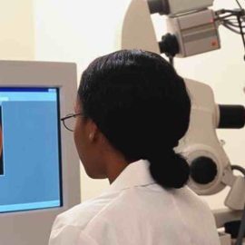
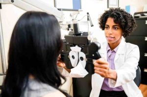
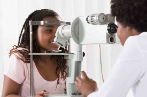
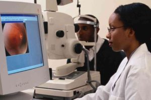
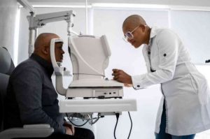
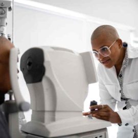
Accurate diagnosis is paramount in guiding treatment decisions and monitoring disease progression. Visual acuity testing, corneal topography, slit-lamp examination, and Pentacam imaging are among the essential diagnostic tools used to evaluate corneal ectasia. These techniques allow ophthalmologists to assess corneal curvature, thickness, and biomechanical properties, aiding in early detection and personalized treatment planning.

Learn about the diagnosis, detection, and treatment of corneal ectasia in Nigeria. This comprehensive guide provides valuable information on identifying and managing this condition effectively.

Symptoms and Clinical Presentation

Patients with corneal ectasia often present with symptoms such as blurred vision, distorted images, and increased sensitivity to light. These visual disturbances can significantly impact daily activities and diminish quality of life. Recognizing the subtle signs of corneal ectasia is essential for timely intervention and preventing irreversible vision loss.

Challenges of Diagnosis in Nigeria

In Nigeria, limited access to advanced diagnostic equipment and specialized ophthalmic services can hinder early detection of corneal ectasia. Additionally, a lack of awareness among both patients and healthcare providers may lead to delays in diagnosis and inadequate management. Addressing these challenges requires a multi-faceted approach, including investment in infrastructure, education, and outreach initiatives.

Treatment Options

The management of corneal ectasia aims to stabilize the cornea, improve visual function, and prevent further deterioration. Spectacles and contact lenses are often the first line of treatment, providing visual correction and symptom relief. For progressive ectasia, interventions such as collagen cross-linking (CXL) and intrastromal corneal ring segments (ICRS) may be recommended to strengthen the corneal tissue and reshape the corneal surface. In severe cases, corneal transplantation may be necessary to restore vision.

Age-related Macular Degeneration (AMD) can impact your vision and ability to drive safely in Nigeria. Learn about the risks and precautions to take while driving with AMD.

Emerging Therapies

Advances in ophthalmic research have led to the development of novel therapies for corneal ectasia. Accelerated cross-linking protocols, which utilize higher energy levels and shorter treatment times, offer the potential for faster recovery and improved outcomes. Additionally, innovative surgical techniques and pharmacological interventions are being explored to address the underlying mechanisms of corneal ectasia and enhance treatment efficacy.

Importance of Patient Education

Patient education plays a crucial role in the management of corneal ectasia, empowering individuals to take an active role in their eye health. It is essential to educate patients about the importance of compliance with treatment regimens, lifestyle modifications, and regular follow-up visits. Support groups and counseling services can provide valuable emotional support and practical advice to patients navigating the challenges of living with corneal ectasia.

Case Studies and Success Stories

Real-life case studies offer insights into the clinical course of corneal ectasia and the impact of various treatment modalities on patient outcomes. By sharing success stories, we can inspire hope and encourage individuals facing similar challenges to seek timely intervention and pursue treatment options that best suit their needs.

Cataract Surgery Following LASIK in Nigeria

Collaborative Efforts in Nigeria

Addressing the complex challenges of corneal ectasia requires collaboration among healthcare professionals, government agencies, and non-profit organizations. Ophthalmological societies play a vital role in promoting education, advocacy, and research in the field of corneal diseases. Government initiatives aimed at expanding access to eye care services and improving the quality of diagnostic and treatment facilities are essential for reducing the burden of corneal ectasia in Nigeria. Partnership with international organizations can facilitate knowledge exchange and resource sharing, ultimately benefiting patients and healthcare providers alike.

Preventive Measures

Preventing corneal ectasia begins with early detection and intervention. Implementing screening programs in schools, communities, and primary care settings can identify at-risk individuals and facilitate timely referral to ophthalmic specialists. Public health campaigns aimed at raising awareness about the signs and symptoms of corneal ectasia can empower individuals to seek prompt medical attention and access appropriate treatment options.

Age-related Macular Degeneration (AMD) can impact your vision and ability to drive safely in Nigeria. Learn about the risks and precautions to take while driving with AMD.

Future Directions in Research

Continued investment in research is essential for advancing our understanding of corneal ectasia and developing innovative therapies. Genetic studies may uncover novel biomarkers for predicting disease progression and identifying individuals at high risk of developing ectatic disorders. Advanced imaging techniques, such as optical coherence tomography (OCT) and high-resolution ultrasound, hold promise for improving the accuracy of diagnosis and monitoring treatment response. Furthermore, the development of targeted pharmacological interventions that modulate corneal biomechanics and cellular metabolism may offer new avenues for personalized treatment approaches.

Conclusion

In closing, the journey through the diagnosis, detection, and treatment of corneal ectasia in Nigeria is not merely a medical endeavor; it is a testament to resilience, hope, and the unwavering commitment to restoring sight and improving lives. Despite the challenges posed by limited resources and awareness, the tireless efforts of dedicated healthcare professionals, coupled with the courage and determination of patients, illuminate a path forward. Every successful diagnosis is a beacon of hope, every treatment a triumph of the human spirit over adversity. As we navigate the complexities of corneal ectasia, let us not forget the faces behind the statistics—the individuals whose lives are forever changed by the transformative power of compassionate care. In Nigeria, as in every corner of the world, the pursuit of sight is a journey worth undertaking, fueled by empathy, solidarity, and the belief that together, we can overcome even the most daunting of obstacles.


Read more

Evaluating the Viability of Cataract Surgery Following LASIK in Nigeria

When it comes to eye health, cataracts are a common concern for many people. In Nigeria, the prevalence of cataracts is on the rise, affecting a significant portion of the population. But what about those who have previously undergone LASIK surgery? Is cataract surgery still a viable option for them? In this blog post, we will explore the viability of cataract surgery following LASIK in Nigeria and delve into the latest advancements in cataract surgery techniques. So, if you’re considering cataract surgery after LASIK or simply curious about the topic, keep reading to learn more.

Cataract Surgery Following LASIK in Nigeria

Prevalence of Cataracts in Nigeria

Cataracts are a common eye condition in Nigeria, affecting a significant portion of the population.

The prevalence of cataracts in Nigeria is increasing due to various factors such as the aging population and lifestyle choices.

Effects of LASIK on Cataract Development

LASIK surgery, a popular refractive procedure, has been extensively studied to assess its impact on cataract development. The findings consistently indicate that LASIK surgery does not significantly increase the risk of developing cataracts.

Multiple research studies have shown that there is no causal relationship between LASIK surgery and the development or progression of cataracts. Patients who have undergone LASIK surgery do not experience a higher incidence or faster progression of cataracts compared to the general population.

This is reassuring for individuals considering LASIK surgery as a vision correction option, especially for those with concerns regarding the potential development of cataracts in the future.


Challenges in Diagnosing Cataracts Post-LASCataract Surgery Following LASIK in NigeriaIK

Diagnosing cataracts post-LASIK can be challenging due to changes in the cornea caused by the surgery. LASIK alters the shape of the cornea, making it difficult to accurately assess the presence and severity of cataracts.

Traditional methods of diagnosing cataracts, such as slit-lamp examination and visual acuity testing, may not provide accurate results in patients who have undergone LASIK. The corneal irregularities caused by LASIK can affect the clarity of the lens in the eye, making it difficult to detect cataracts.

Specialized imaging techniques, such as optical coherence tomography (OCT) and specular microscopy, may be necessary to accurately diagnose cataracts in patients who have undergone LASIK. These imaging technologies provide detailed images of the cornea and can help ophthalmologists identify and evaluate cataracts.

Ophthalmologists need to be aware of the challenges in diagnosing cataracts post-LASIK and utilize advanced imaging techniques to ensure accurate diagnosis and appropriate treatment options for patients.

Advancements in Cataract Surgery Techniques

Over the years, significant advancements have been made in cataract surgery techniques, making the procedure safer and more effective for patients in Nigeria. These advancements have revolutionized the way cataracts are treated, leading to improved surgical precision and outcomes.

One notable advancement is the introduction of femtosecond laser-assisted cataract surgery. This innovative technique utilizes a laser to perform key steps of the surgery, such as creating precise incisions and fragmenting the cataract for easier removal. By utilizing laser technology, surgeons can enhance the accuracy and predictability of the procedure, resulting in better visual outcomes for patients.

Additionally, new types of intraocular lenses (IOLs) have been developed to address specific visual needs and enhance postoperative visual acuity. These advanced IOLs include multifocal and toric lenses, which can correct both distance and near vision or astigmatism, respectively. By choosing the right IOL based on the patient’s lifestyle and visual requirements, surgeons can optimize the visual outcomes of cataract surgery.

Advancements in surgical techniques and technology have also led to reduced surgical time and faster recovery for patients. With improved microsurgical tools and techniques, surgeons can perform cataract surgery with increased safety and efficiency. As a result, patients experience less postoperative discomfort and enjoy the benefits of improved vision sooner.

As technology continues to advance, it is expected that cataract surgery techniques will further evolve. The integration of artificial intelligence (AI) into surgical planning and execution may provide even greater precision and customization in cataract surgery. AI algorithms can analyze patient data and optimize surgical parameters to maximize visual outcomes.

Advancements in cataract surgery techniques have significantly improved the safety, efficacy, and predictability of the procedure. Patients in Nigeria can benefit from these advancements, which have led to better visual outcomes, faster recovery, and an overall improved quality of life post-surgery.

Cataract Surgery Following LASIK in Nigeria

Cataract Surgery Success Rates in Nigeria

Cataract surgery success rates in Nigeria are comparable to international standards. The majority of patients experience improved vision and quality of life after cataract surgery.

Mitigating Risks in Cataract Surgery after LASIK

Preoperative planning and careful evaluation of the patient’s corneal health can help mitigate risks in cataract surgery after LASIK. It is important to assess the stability of the cornea and the quality of vision before proceeding with cataract surgery.

Specialized imaging techniques, such as optical coherence tomography (OCT) and corneal topography, can provide valuable information about the cornea’s thickness, shape, and integrity. These tests can help identify any irregularities or abnormalities that may affect the surgical outcome.

Additionally, using advanced technology and techniques can minimize potential complications. For instance, utilizing the femtosecond laser in cataract surgery can enhance precision and reduce the risk of corneal damage. It allows for the creation of precise incisions, capsulotomies, and fragmentation of the cataract, improving overall surgical outcomes.

Regular follow-up visits and thorough postoperative care are also crucial in identifying and managing any potential complications or changes in visual acuity. Patients should be educated about the importance of compliance with postoperative medications and precautions to ensure optimal healing and recovery.

Choosing the Right Intraocular Lens for Cataract Surgery

When undergoing cataract surgery, choosing the right intraocular lens (IOL) is crucial for achieving optimal visual outcomes. Several factors should be considered when selecting the lens:

Patient’s Lifestyle

The patient’s lifestyle plays an important role in determining the type of IOL that will best suit their needs. For example, active individuals who participate in sports or outdoor activities may benefit from a multifocal or toric IOL that provides enhanced visual clarity at various distances.

Visual Needs

Understanding the patient’s visual needs is essential when choosing an IOL. Some patients may prioritize distance vision, while others may require improved near vision for activities such as reading or using digital devices. Different types of IOLs, such as monofocal, multifocal, or accommodating lenses, can address specific visual needs.

Budget

The cost of the IOL is another important consideration. While advanced premium IOLs offer additional benefits, they may come at a higher price. It is essential to discuss the available options with the patient, taking into account their budget.

By carefully considering these factors, the ophthalmologist can help the patient make an informed decision regarding the type of intraocular lens that will best meet their needs and preferences.

Cataract Surgery Following LASIK in Nigeria

Recovery and Rehabilitation after Cataract Surgery

After cataract surgery, patients typically experience a quick recovery, with improved vision noticeable within a few days. However, it is important to follow postoperative care instructions to ensure optimal healing and rehabilitation.

During the initial recovery period, it is common to experience mild discomfort, itching, foreign body sensation, or light sensitivity. These symptoms usually subside within a few days, but it is essential to avoid rubbing or putting pressure on the operated eye.

Eye drops prescribed by the surgeon should be used as directed to help prevent infection and reduce inflammation. It is crucial to maintain good hygiene and avoid dusty or smoky environments during the healing process.

Rehabilitation exercises, such as daily gentle eye massages, may be recommended to promote healing and reduce the risk of developing scar tissue. These exercises should be performed under the guidance of the surgeon or a trained professional.

Follow-up appointments with the surgeon are necessary to monitor progress and ensure there are no complications. It is important to attend these appointments and inform the surgeon if any unusual symptoms or changes in vision occur.

Most patients experience a significant improvement in vision and quality of life after cataract surgery. However, it is important to note that everyone’s healing process may vary. It may take several weeks or even months for vision to stabilize completely.

Patient Satisfaction with Cataract Surgery in Nigeria

High levels of patient satisfaction have been reported in Nigeria following cataract surgery. Patients have reported significant improvement in visual acuity and overall quality of life.

Cataract Surgery Following LASIK in Nigeria

Future Directions in Cataract Surgery Post-LASIK

As technology continues to advance, there are several exciting developments in the field of cataract surgery post-LASIK:

  1. Laser-assisted Cataract Surgery: The use of lasers in cataract surgery is gaining momentum. Femtosecond laser-assisted cataract surgery offers greater precision and potentially faster recovery times. Ongoing research aims to optimize the benefits of laser technology in cataract surgery post-LASIK.
  2. Intraoperative Wavefront Aberrometry: Wavefront aberrometry measures and corrects optical imperfections during cataract surgery, resulting in improved visual outcomes. Studies are exploring the utility of this technology in patients who have previously undergone LASIK.
  3. Artificial Intelligence (AI) in Cataract Surgery: AI has the potential to revolutionize cataract surgery. Machine learning algorithms can assist surgeons in preoperative planning and improve surgical accuracy. Continued research may uncover new AI-based techniques specific to cataract surgery post-LASIK.
  4. Hybrid Surgeries: Combining cataract surgery with refractive procedures like LASIK or PRK is an area of ongoing investigation. These hybrid surgeries aim to correct both cataracts and refractive errors simultaneously, providing optimal visual outcomes for patients.

With these advancements and ongoing research, the future of cataract surgery post-LASIK looks promising. Surgeons in Nigeria and beyond are dedicated to staying at the forefront of these developments to provide the best possible care for their patients.

Conclusion

In conclusion, cataract surgery following LASIK in Nigeria is a viable option for patients seeking improved vision and quality of life. Despite the challenges in diagnosing cataracts post-LASIK, advancements in imaging techniques have made accurate diagnosis possible. The success rates of cataract surgery in Nigeria are comparable to international standards, and patients have reported high levels of satisfaction with the procedure. Mitigating risks through careful preoperative planning and utilizing advanced technology can further enhance surgical outcomes. Additionally, ongoing research and advancements in cataract surgery techniques offer promising future directions for improved outcomes. Overall, those considering cataract surgery in Nigeria following LASIK can have confidence in the safety and effectiveness of the procedure.


Read more

Understanding Retinal Disorders: A Comprehensive Guide for Nigerians

Retinal disorders encompass a range of conditions that affect the delicate tissue at the back of the eye responsible for vision. These disorders can have a significant impact on visual acuity and overall eye health. This comprehensive guide aims to provide Nigerians with a better understanding of common retinal disorders, their causes, symptoms, diagnosis, and available treatment options.

The retina is the light-sensitive layer of tissue that lines the back of the eye. It converts light into electrical signals that are sent to the brain through the optic nerve, where they are processed into images. The retina is essential for vision, and any damage or disease that affects it can lead to vision loss or blindness.

Retinal disorders are a group of conditions that affect the retina and its function. They can be caused by various factors, such as aging, genetics, infections, injuries, diabetes, hypertension, or other systemic diseases. Some of the common retinal disorders include:

1. Age-related macular degeneration (AMD): This is a degenerative condition that affects the macula, the central part of the retina that is responsible for sharp and detailed vision. AMD causes gradual loss of central vision, making it difficult to read, drive, or recognize faces. AMD is the leading cause of blindness among older adults in Nigeria and worldwide.

2. Diabetic retinopathy (DR): This is a complication of diabetes that damages the blood vessels in the retina. DR can cause bleeding, swelling, or leakage of fluid in the retina, leading to blurred or distorted vision. DR is the leading cause of blindness among working-age adults in Nigeria and worldwide.

3. Retinitis pigmentosa (RP): This is a hereditary disorder that affects the photoreceptors, the cells in the retina that detect light. RP causes progressive loss of peripheral vision and night vision, resulting in tunnel vision or blindness. RP is one of the leading causes of bilateral blindness in Nigeria.

4. Retinal detachment: This is a medical emergency that occurs when the retina separates from the underlying layer of tissue. Retinal detachment can cause flashes, floaters, or a curtain-like shadow in the visual field. If not treated promptly, retinal detachment can lead to permanent vision loss.

5. Retinal vein occlusion (RVO): This is a blockage of one of the veins that drain blood from the retina. RVO can cause sudden or gradual loss of vision, depending on the location and extent of the blockage. RVO can also lead to complications such as macular edema (swelling of the macula) or neovascularization (growth of abnormal blood vessels).

6. Macular hole: This is a small break in the macula that causes a central blind spot or distortion in vision. Macular hole can be caused by aging, trauma, inflammation, or vitreomacular traction (pulling of the vitreous gel on the macula).

7. Parafoveal telangiectasia: This is a rare condition that affects the small blood vessels around the macula. Parafoveal telangiectasia causes leakage of fluid or blood in the macula, leading to blurred or distorted vision.

Retinal disorders encompass a range of conditions that affect the delicate tissue at the back of the eye responsible for vision. These disorders can have a significant impact on visual acuity and overall eye health. This comprehensive guide aims to provide Nigerians with a better understanding of common retinal disorders, their causes, symptoms, diagnosis, and available treatment options.

The diagnosis and treatment of retinal disorders depend on the type and severity of the condition. Some of the common diagnostic tests include:


1. Visual acuity test: This measures how well you can see at different distances.

2. Fundus examination: This uses an instrument called an ophthalmoscope to examine the back of your eye, including the retina and optic nerve.

3. Optical coherence tomography (OCT): This uses light waves to create detailed images of your retina and its layers.

4. Fundus fluorescein angiography (FFA): This uses a dye that is injected into your arm and travels to your eye. A special camera then takes pictures of your retina as the dye highlights its blood vessels.

5. Electroretinography (ERG): This measures the electrical activity of your retina in response to light stimuli.

retinal disorders

Some of the common treatments for retinal disorders include:


1. Medications: These include eye drops, injections, or oral drugs that aim to reduce inflammation, swelling, bleeding, or abnormal blood vessel growth in the retina.

2. Laser therapy: This uses a beam of light to seal leaking or abnormal blood vessels, shrink tumors, or create scars to prevent retinal detachment.

3. Vitrectomy: This is a surgical procedure that removes some or all of the vitreous gel from your eye. Vitrectomy can help treat retinal detachment, macular hole, vitreomacular traction, or severe bleeding in the eye.

4. Scleral buckle: This is a surgical procedure that places a silicone band around your eye to push it against the detached retina and reattach it.

5. Pneumatic retinopexy: This is a surgical procedure that injects a gas bubble into your eye to push against the detached retina and reattach it.

6. Retinal implant: This is a device that is surgically implanted into your eye to replace damaged photoreceptors and restore some vision. Retinal implants are still experimental and only available for certain types of retinal disorders.


The prevention and management of retinal disorders depend on the underlying cause and risk factors. Some of the general tips include:

1. Regular eye check-ups: This can help detect and treat retinal disorders early, before they cause irreversible vision loss.

2. Control of blood sugar, blood pressure, and cholesterol levels: This can help prevent or delay the onset of diabetic retinopathy or retinal vein occlusion.

3. Quitting smoking and limiting alcohol intake: This can help reduce the risk of age-related macular degeneration or other retinal diseases.

4. Eating a balanced diet rich in antioxidants, vitamins, and minerals: This can help protect your retina from oxidative stress and inflammation.

5. Wearing sunglasses and hats: This can help protect your retina from harmful ultraviolet rays and blue light.

6. Avoiding eye injuries and infections: This can help prevent trauma or inflammation that can damage your retina.

7. Seeking immediate medical attention for any sudden or severe vision changes: This can help prevent permanent vision loss from retinal detachment or other emergencies.

Conclusion

Retinal disorders are a major cause of visual impairment and blindness in Nigeria and worldwide. They can affect anyone at any age, but some people are more prone to them than others. By understanding the types, causes, symptoms, diagnosis, treatment, and prevention of retinal disorders, you can take better care of your eyes and your vision.

Read more